Table of Contents
ToggleMonopolar Laparoscopic Instruments: Master Safety, Avoid Costly Burns
Monopolar electrosurgery is a cornerstone of modern laparoscopic surgery, offering unparalleled precision in cutting and coagulating tissue. Its efficiency in achieving hemostasis and facilitating dissection has revolutionized minimally invasive procedures. However, the energy that grants this surgical advantage carries a significant risk: unintended thermal injury. “Costly burns,” whether to the patient at the active site, at remote sites, or even to the surgical team, represent one of the most feared complications of monopolar energy. This detailed article aims to equip surgeons and the entire perioperative team with the knowledge and practical strategies to “master safety” and consistently avoid these devastating complications.
The Power of Monopolar Energy: A Double-Edged Scalpel
At its core, monopolar electrosurgery works by concentrating high-frequency electrical current at the tip of an active electrode. This current then passes through the patient’s body to a large, dispersive return electrode (often called a grounding pad) placed on the patient’s skin, completing an electrical circuit back to the electrosurgical unit (ESU). The high current density at the active electrode causes rapid heating and vaporization of tissue (cutting) or desiccation and coagulation (hemostasis).
Benefits of Monopolar Electrosurgery in Laparoscopy:
- Precise Cutting: Allows for clean, controlled incisions through tissue.
- Effective Coagulation: Rapidly seals small blood vessels, minimizing blood loss and improving visibility.
- Versatility: Can be integrated into various instruments like dissectors, scissors, hooks, and spatulas.
- Reduced Operative Time: Efficient hemostasis and cutting can significantly shorten procedure duration.
- Cost-Effectiveness: Often more economical than some alternative energy sources.
However, the very pathway of the current through the patient’s body inherently carries risks. Understanding these risks is the first step towards mitigation.
The Anatomy of a Burn: Understanding the Mechanisms of Injury
Monopolar electrosurgical burns primarily arise from uncontrolled or unintended current pathways. These can be broadly categorized into:
1. Direct Application Burns: The Obvious Risk
This occurs when the active electrode makes direct, unintended contact with non-target tissue, often out of the surgeon’s direct line of sight.
- Mechanism: Inadvertent activation of the ESU foot pedal while the instrument is near or touching a vital structure (e.g., bowel, ureter, vessel) not intended for thermal effect. It can also happen when the activated tip brushes against an organ during insertion or withdrawal through a trocar.
- Prevention:
- “See what you heat”: Always ensure the active tip is fully visible and in direct contact with the target tissue before activation.
- Controlled Activation: The surgeon should be the sole activator of the ESU. Foot pedals should be placed to prevent accidental activation.
- Instrument Out When Not In Use: When not actively using the monopolar instrument, remove it from the patient or place it in a dry, rigid, insulated holder. Never leave it in contact with patient tissue.
- Short, Intermittent Activations: To achieve the desired effect, use the lowest effective power setting and short bursts of energy. Avoid prolonged activation.
2. Return Electrode (Dispersive Pad) Burns: The “Grounding Pad” Hazard
These burns occur when the current returning to the ESU through the dispersive pad is not adequately dispersed, leading to a high current density at the skin-pad interface.
- Mechanism:
- Inadequate Contact: The pad is not fully adhered to the skin, has air pockets, or becomes dislodged during surgery.
- Reduced Surface Area: The Pad is too small for the patient’s size or power setting.
- Conductive Bridging: Fluid (e.g., saline, blood) pools under the pad, creating a smaller, more conductive pathway for the current.
- Placement Over Bony Prominences/Scar Tissue: These areas have higher impedance, leading to increased heat generation.
- Old/Damaged Pad: Adhesive or conductive gel integrity may be compromised.
- Prevention:
- Proper Placement: Apply the dispersive electrode to a large, well-vascularized muscle mass, free of hair, scars, bony prominences, or implants.
- Full Adherence: Ensure the pad’s entire surface is in firm, uniform contact with the skin. Avoid wrinkles or creases.
- Monitor Contact Quality (CQM): Utilize ESUs equipped with Contact Quality Monitoring (CQM) systems, which continuously monitor the impedance at the pad and alarm if contact is compromised.
- Avoid Fluid Pooling: Ensure the surgical field is dry, especially around the pad.
- Regular Checks: Re-check pad position and adherence if the patient’s position is changed intra-operatively.
3. Insulation Failure: The Silent Threat
This is arguably the most insidious cause of laparoscopic thermal injury, as it often occurs out of the surgeon’s direct vision.
- Mechanism: Damage to the insulating coating of the active electrode allows current to leak from the instrument’s shaft to unintended tissues. This can occur due to:
- Repetitive Use: Wear and tear from repeated passage through trocars, particularly with sharp edges or during instrument rotation.
- Cleaning/Sterilization: Harsh chemicals, high temperatures, or mechanical damage during reprocessing.
- Manufacturing Defects: Rare, but possible.
- Small Defects, High Risk: Smaller insulation defects create higher current density at the point of leakage, leading to more severe localized burns.
- Prevention:
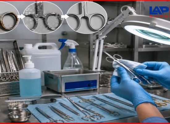
- Meticulous Inspection: Visually inspect every reusable monopolar instrument before and after every use for any signs of insulation damage (cracks, nicks, discoloration). A magnifying glass can be helpful.
- Discard Damaged Instruments: Any instrument with insulation damage must be immediately removed from service and repaired or discarded.
- Active Electrode Monitoring (AEM): Utilize instruments and ESUs with AEM technology. AEM systems incorporate a protective shield around the active electrode that detects stray current and either redirects it back to the ESU or alarms and shuts down the generator, preventing a burn.
- Appropriate Trocar Size: Use a trocar size appropriate for the instrument diameter to minimize friction and damage during insertion and withdrawal.
- Single-Use Instruments: While more costly, disposable instruments eliminate the risk of instrument degradation from reprocessing.
4. Capacitive Coupling: The “Invisible Current”
This phenomenon occurs when electrical current is transferred from the active electrode, through intact insulation, to an adjacent conductive material (e.g., another metal instrument, a metal trocar cannula) without direct contact.
- Mechanism: A high-frequency alternating current in the active electrode creates an electrical field that can induce a current in nearby conductive objects. This induced current can then flow to patient tissue, particularly if the secondary conductor is “isolated” (e.g., a metal instrument inside a plastic trocar cannula) or if the tissue contact area is small, leading to a high current density and burn.
- Prevention:
- All-Metal or All-Plastic Cannulas: Avoid “hybrid” trocar systems (plastic outer sleeve with a metal inner channel) that can trap capacitively coupled energy. All-metal cannulas safely dissipate the energy to the abdominal wall.
- Active Electrode Monitoring (AEM): AEM systems are highly effective at preventing capacitive coupling by diverting stray energy back to the generator.
- Minimize “Open Air” Activation: Activating the ESU while the instrument is not in contact with tissue can increase voltage and thus the risk of capacitive coupling. Touch the tissue before activating.
- Lowest Effective Power: Use the lowest power settings necessary to achieve the desired tissue effect. Higher voltages increase capacitive coupling risk.
- Short Bursts: Reduce activation time.
5. Direct Coupling: The “Spark Jump”
Direct coupling occurs when an active electrode (while activated) inadvertently touches another metal instrument or conductive object within the surgical field that is in contact with non-target tissue.
- Mechanism: The electrical current “jumps” from the active electrode to the adjacent conductive instrument, and then flows through that instrument to the tissue it’s touching, creating a burn. This is particularly dangerous as the burn can occur far from the surgeon’s immediate view.
- Prevention:
- Maintain Visual Control: Always keep the active electrode and the area around its tip in constant view on the monitor.
- Avoid “Sword Fighting”: Do not allow instruments to cross or touch each other while the ESU is activated.
- Adequate Working Space: Ensure sufficient space between instruments and tissues to prevent accidental contact.
- Retract Non-Target Tissue: Use other instruments to retract and isolate non-target structures from the active electrode.
Essential Safety Protocols and Best Practices
Beyond understanding the mechanisms, implementing a robust safety protocol is paramount.
Pre-Operative Checklist:
- Patient Assessment: Identify patients with pacemakers, ICDs, or other electronic implants. Consult cardiology if monopolar use is necessary; these devices may need to be interrogated or deactivated.
- Skin Integrity: Inspect the patient’s skin for lesions, tattoos, scars, or excessive hair before applying the return electrode.
- Return Electrode Placement: Ensure proper placement, full adherence, and appropriate size for the patient and anticipated power settings.
- Instrument Inspection: Visually inspect all reusable monopolar instruments for insulation defects, damage, or wear.
- Cable Integrity: Check all ESU cables for frays, cuts, or damaged connectors.
- ESU Settings: Confirm the ESU is set to the correct mode (cut, coag, blend) and the lowest effective power for the specific procedure.
- Flammable Materials: Ensure all flammable prep solutions have thoroughly dried, and remove any alcohol-based materials from the sterile field.
Intra-Operative Guidelines:
- Visual Confirmation: Never activate the ESU unless the active electrode tip is in clear view and contact with the intended target tissue.
- Lowest Effective Power: Use the lowest power setting that achieves the desired tissue effect. Avoid excessive power.
- Short Bursts of Energy: Activate the ESU in short, controlled bursts rather than prolonged activation. This minimizes thermal spread.
- Tissue Tension: Apply slight tension to the tissue being dissected or coagulated; this can improve the effectiveness of the energy.
- Clean Instrument Tips: Charred tissue on the active electrode tip increases impedance and requires higher power, leading to greater thermal spread. Clean the tip frequently.
- Avoid Activation in Air: Do not activate the ESU in an open circuit (i.e., in the air or without tissue contact), as this can increase voltage and promote capacitive coupling.
- Insulated Holster: When not in active use, place the active electrode in an insulated safety holster to prevent accidental activation or contact.
- Constant Communication: Maintain open communication with the surgical team, particularly the ESU operator (if not the surgeon).
- Awareness of “Out of View” Areas: Always be mindful of instruments, tissue, and vital structures that may be out of direct laparoscopic view, especially when activating energy.
- Post-Activation Cooling: Recognize that instruments retain heat after activation. Avoid touching non-target tissue immediately after an energy burst.
Post-Operative Considerations:
- Skin Inspection: Re-inspect the return electrode site and other potential remote burn sites for any signs of injury.
- Documentation: Document ESU settings, return electrode type and placement, and any concerns or incidents.
- Patient Education: Advise patients about potential delayed complications (e.g., abdominal pain, fever) that could indicate an unseen thermal injury to the bowel or other organs.
Types of Monopolar Laparoscopic Instruments
Monopolar energy can be delivered through various laparoscopic instruments, each designed for specific surgical tasks:
- Monopolar Hooks: Used for precise dissection and coagulation of small vessels. Available in J-hook, L-hook, and spatula designs.
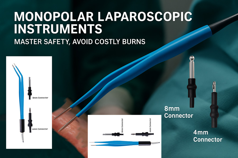
- Monopolar Maryland/Dissecting Forceps: Versatile instruments for grasping, blunt dissection, and coagulation. The curved jaws allow for precise application of energy.
- Monopolar Scissors: For cutting tissue with simultaneous coagulation. Available in curved or straight designs.
- Monopolar Spatulas/Needles: Used for fine dissection and pinpoint coagulation.
- Monopolar Electrodes (Ball Tip, Rollerball): Primarily for broad surface coagulation or desiccation.
Common Monopolar Burn Mechanisms and Prevention Strategies
| Burn Mechanism | Description | Key Prevention Strategies |
|---|---|---|
| Direct Application | Active electrode contacts non-target tissue due to inadvertent activation. | “See what you heat,” controlled activation, instrument out when not in use, short activations, lowest effective power. |
| Return Electrode | Inadequate current dispersion at the dispersive pad site. | Proper placement of muscle mass, full adherence, CQM monitoring, avoiding bony prominences/scars, and avoiding fluid pooling. |
| Insulation Failure | Current leaks through damaged insulation on the instrument shaft. | Meticulous pre-use inspection, discard damaged instruments, Active Electrode Monitoring (AEM), appropriate trocar size, proper handling/reprocessing. |
| Capacitive Coupling | Induced current from active electrode to adjacent conductor (intact insulation). | All-metal or all-plastic cannulas, AEM technology, minimize “open air” activation, and the lowest effective power. |
| Direct Coupling | The active electrode directly contacts another conductive instrument (e.g., a scope). | Maintain visual control, avoid “sword fighting,” adequate working space, and retract non-target tissue. |
The Role of Technology: Active Electrode Monitoring (AEM)
Active Electrode Monitoring (AEM) represents a significant advancement in monopolar electrosurgery safety. AEM systems incorporate a conductive shield around the active electrode’s insulation. This shield is connected to the ESU’s return circuit.
- How it Works: If there is insulation failure or capacitive coupling, stray current is detected by the shield. Instead of arcing to adjacent tissue, the current is safely shunted back to the ESU. In many systems, an alarm sounds, and the ESU may even shut down, alerting the surgeon to the potential hazard before a burn occurs.
- Benefits: AEM technology significantly reduces the risk of undetected thermal injuries caused by insulation failure and capacitive coupling, providing an invaluable layer of safety in laparoscopic procedures. While not a substitute for vigilant technique and instrument inspection, it offers an important safety net.
The Human Factor: Training and Teamwork
Ultimately, safety in monopolar electrosurgery hinges on the competence and vigilance of the entire surgical team.
- Comprehensive Training: All perioperative personnel involved in electrosurgery must receive thorough training on the principles of electrosurgery, specific ESU operation, instrument handling, and safety protocols.
- Team Communication: A culture of open communication where any team member can voice a concern about instrument integrity or potential hazards is crucial.
- Continuous Education: Regular refreshers and updates on best practices and new technologies are essential.
- Incident Reporting: A robust system for reporting and analyzing electrosurgical incidents (even “near misses”) allows for learning and improvement.
Monopolar laparoscopic instruments are powerful tools that have transformed surgical care. However, their power demands unwavering respect and meticulous adherence to safety principles. “Mastering safety” in monopolar electrosurgery is not merely about avoiding “costly burns” but about cultivating a deep understanding of energy principles, rigorous instrument management, and a culture of proactive vigilance within the operating room. By embracing comprehensive training, implementing stringent safety protocols, and leveraging technological advancements like Active Electrode Monitoring, surgeons and their teams can continue to harness the immense benefits of monopolar energy while ensuring the highest level of patient safety, truly avoiding costly burns and devastating complications.
For more information, feel free to contact the Lapex Surgical support team.